![[Logo] Instituto Superior
Técnico](img/wwwist.gif)
Aviso:Se está a ler esta mensagem, provavelmente, o browser que utiliza não é compatívelcom os "standards" recomendados pela W3C.Sugerimos vivamente que actualize o seu browser para ter uma melhor experiênciade utilização deste "website".Mais informações em webstandards.org.
Warning: If you are reading this message, probably, your browser is not compliant with the standards recommended by the W3C. We suggest that you upgrade your browser to enjoy a better user experience of this website.More information on webstandards.org.

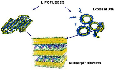
Lipid/DNA complexesGene therapy offers promise for the treatment of disease through the use of DNA based vectors that allow targeting, delivery of DNA to cells and expression of the gene. Among the non-viral vectors, cationic liposomes seem to be the most widely used DNA delivery system. Despite low transfection efficiencies, they show nonimmunogenicity, low toxicity and possibility of large scale production. Many efforts have been made to fully characterize cationic liposome-DNA complexes (lipoplexes), because it is the only way to understand, improve and control the transfection efficiency of these non-viral based vectors. In 1987, it was reported for the first time that plasmid DNA and cationic liposomes aggregate due to electrostatic attractive forces and origin small complexes able to transfer DNA to the cells. In the late 90's, electron microscopy and X-ray diffraction used in parallel revealed a multilamellar structure of lipid bilayers with sandwiched DNA, with a constant interlayer spacing invariant with the charge ratio, and depending on cationic liposomes formulations (Figure 1).
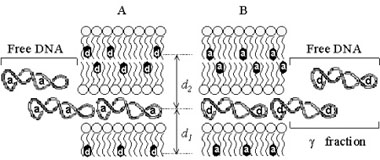
In our lab, we developed FRET models assuming a multilamellar lipoplex arrangement (Figure 2). The application of these models allows the determination of the distance between the fluorescent intercalator on the DNA and a membrane dye on the lipid, and/or the evaluation of encapsulation efficiencies of this liposomal vehicle.
|
|||||||