![[Logo] Instituto Superior
Técnico](img/wwwist.gif)
Aviso:Se está a ler esta mensagem, provavelmente, o browser que utiliza não é compatívelcom os "standards" recomendados pela W3C.Sugerimos vivamente que actualize o seu browser para ter uma melhor experiênciade utilização deste "website".Mais informações em webstandards.org.
Warning: If you are reading this message, probably, your browser is not compliant with the standards recommended by the W3C. We suggest that you upgrade your browser to enjoy a better user experience of this website.More information on webstandards.org.

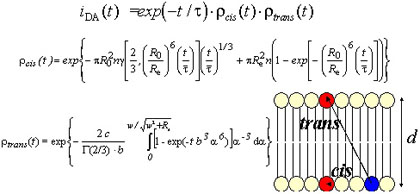
Fluorescence Resonance Energy Transfer (FRET)A particularly powerful fluorescence technique to probe membrane organization and lipid-protein interaction is fluorescence resonance energy transfer (FRET; Theodore Förster, 1949). The rate of energy transfer between a donor molecule, with fluorescence lifetime τ, and an acceptor molecule, separated by a distance R, is given by  where R0 is the critical distance, which can be calculated from 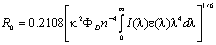 where in turn κ2 is the orientation factor, ΦD is the donor quantum yield in the absence of acceptor, n is the refractive index, λ is the wavelength, I(λ) is the normalized donor emission spectrum, and ε(λ) is the acceptor molar absorption spectrum. In the latter equation, if the λ units used are nm, the calculated R0 has Å units. If each donor senses a single acceptor in its vicinity, and the donor-acceptor distance is the same for all pairs, then FRET is easily used as a "spectroscopic ruler" to measure distances in the 1-10 nm range. In case the donor molecules are surrounded by a distribution of acceptors, the decay law becomes complex (Figure 1), and depends on the acceptor concentration and distribution.
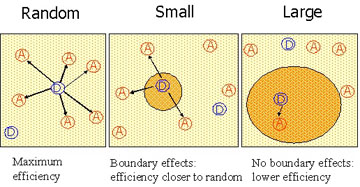
Membrane phase separation leads to partition of both donor and acceptor probes between the two phases. In general, the donor fraction and fluorescence properties will be different in the two phases, as will be the local acceptor concentrations. Analysis of time-resolved donor fluorescence in presence and absence of acceptor allows the recovery of all these parameters, and, from deviations to the theoretical expectations, information about domain size can be inferred (Figure 2).
Regarding lipid-protein interaction, FRET is helpful in quantification of lipid-protein selectivity, determining the transverse location of the peptide/protein fluorophore, and probing the peptide/protein aggregation state, among other other important questions. |
|||||||